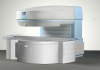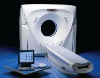

Mx -8000 Quad 4-slice CT, Fully refurbished, with Brandnew Tube. Provide Worldwide Installation service. Turn-key Project.
Mx8000 IDT Quad, 4-Slice Ultra fast CT System
Model: MX-8000 (4 slices) fully refurbished and tested in 2012
The Mx-8000 is a high power, ultrahigh resolution premium volumetricCT scanner. It is built on an Ultra Fast (0.75 sec.), multi-slice platformproviding comprehensive clinical solutions in CT at a significantly loweroperation cost.
Mx8000 with breakthrough True Capture Technologies (TCT).
Includes:
Up to 100 seconds of continuous multi-slice spiral capability0.75 second full 360° scan time Temporal resolution as low as 450 msecPatented DFS sampling providing the worlds highest in-plane resolution 24 lp/cmUltra fast, (0.85 Sec) Sub second axial and spiral reconstruction packageBuilt with over a decade of multi-slice experience, The Mx-8000 proprietary spiral interpolation and reconstruction techniques increase multi-slice coverage while maintaining image quality.
ESP II (Easy Scan Planning) software with specialized viewing modes is specifically designed for multi-slice workflow and virtually eliminates technologist learning curves, providing the ultimate productivity tool and gateway to new CT applications.
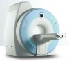
Mx-8000 System components:
Gantry
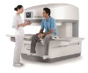
Patient table
Power distribution cabinet
Cooling unit- A/W
Mx-8000 operating, viewing and processing console
High-speed computing and display system
Key System Highlights:
· Ultra Fast, MultiSlice, Spiral Scanner by design
· AsymmetrixTM Philips Patented variable wide area detector providing optimal dose efficiency
· ExcelleratorTM 200 Mbit/second Ultrahigh speed multi-slice data acquisition system
· On-board 60 kW, high frequency, high-voltage generator
· Low-voltage Slip-Ring
· 6.5 MHU high power X-ray tube
· Dynamic Focus System (DFS) doubles data density providing up to 24 lp/cm ultra-high spatial resolution, in axial and spiral scanning
· SubSecond scan mode: 0.5 sec for full 360° scan
· 36 GB raw data storage memory
· 0.85 sec reconstruction time for a large FOV image
· Reconstruction matrices of 340, 512.
· Powerful Silicon Graphics R5000SC RISC host computer with multitasking graphic user interface and ESP II Software
· Brand New 19”, Philips high resolution LCD, color monitor
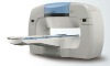
· Patient couch with 1570 mm scannable range
· Gantry and table controls conveniently located on both sides of the gantry and on the operator console
· Large 50 cm field-of-view (FOV) inside a wide, flared
· 70 cm aperture
· Multiple-Slice sequential axial scanning of up to 4 simultaneous slices
· Multiple-Slice, volumetric spiral scanning:up to 100 sec continuous, multiple and bi-directional acquisitions.
· DICOM 3.0 compliant image format, archive and networking
· ESP II Software packages and software license
MxView Diagnostic workstation
The MxView independent multimodality diagnosticworkstation provides quick processing, analysis, manipulation, display,filming, storage and retrieval of images from different imaging modalities.
MxView features: * Cutting-edge clinical image processing
* Real time and user-friendly operation
* Universal connectivity
* Full DICOM 3.0 compliance
* Standard Unix-based workstation
* Windows-like, mouse driven user-interface
* Fast Silicon Graphics workstation based on a RISC processor
* 512 GB RAM
* 1280 x 1024 pixels display on a high resolution color monitor
* 9 GB hard-disk for storage of up to 18,500 (512 x 512 matrix) images (CT9229)
* Post-processing and enhanced clinical applications:
* MPR(Real-time multiplanar Reformatting) of images into any user-defined linear or curved planes
* 3DSSD (Shaded Surface Display ) of up to 15 separate tissues or organs with real-time manipulations and cutting
* VOYAGER-VirtualEndoscopy to provide an interactive exploration of patient anatomy from inside.
* 4D-AngioVolume Rendering to reproduce the whole tissues and organs volume.
* DentalCT for producing panoramic and cross-sectional cuts through the Mandible or Maxilla from CT images
* Q-CTA is atool kit for taking quantitative measurements of anatomic structures, includingvasculature from 2-D, 3-D or 4-D Angio volume-rendering image.
* Time Lapseapplication for graphic display of MRI or CT pixel values vs. time to analyze the uptake and perfusion of contrast media with time.
* MIP
* Combine Images
* Master Look
* MRI Normalization
Mx8000 IDT Quad 4-Slice CT Scanner